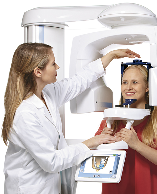
In recent years, advancements in dental imaging technology have significantly transformed the field of dentistry. One of the most impactful innovations is the emergence of 3D dental X-rays, which provide comprehensive views of dental structures, facilitating better diagnosis and treatment planning. Unlike traditional 2D X-rays, 3D dental X-rays offer multi-dimensional images that enhance the accuracy of detecting dental issues. This article provides an in-depth exploration of 3D dental X-rays, covering their benefits, functionality, and applications within the dental industry.
Understanding 3D Dental X-rays
3D dental X-rays, also known as cone beam computed tomography (CBCT), utilize a rotating imaging machine that captures data from multiple angles to create a three-dimensional image of the patient’s teeth, soft tissues, nerve pathways, and bone. This technology is particularly beneficial for diagnosing complex dental conditions and planning intricate procedures such as implant placement, root canal treatments, and orthodontic assessments. Read more about this topic.
Benefits of 3D Dental X-rays
- Enhanced Image Quality: Provides detailed images that allow for more precise diagnosis and treatment planning.
- Comprehensive View: Offers a complete view of the dental structure, including hard-to-see areas like roots and bone structures.
- Reduced Radiation Exposure: Utilizes a lower dose of radiation compared to conventional CT scans.
- Quick and Non-invasive: The imaging process is fast, typically completed within a single session, reducing the need for multiple appointments.
These advantages significantly improve patient care by enabling dentists to make informed decisions. Learn more in this detailed guide.
Applications of 3D Dental X-rays
3D dental X-rays are versatile and applicable across various dental disciplines. Some key applications include:
Implant Dentistry
They assist in accurately planning the placement of dental implants by providing a clear view of the jawbone and surrounding areas, ensuring optimal positioning and integration. Explore further insights here.
Orthodontics
For orthodontists, 3D images offer a detailed analysis of tooth alignment and jaw structure, which is crucial for devising effective treatment plans and monitoring progress.
Endodontics
In endodontics, 3D dental X-rays help in diagnosing root fractures, canal structures, and periapical lesions, leading to more successful root canal treatments. Find additional information here.
Oral Surgery
Surgeons utilize this technology to plan complex surgeries, such as wisdom teeth extraction and corrective jaw surgery, minimizing risks and enhancing surgical outcomes.
How 3D Dental X-rays Work
The process of capturing 3D dental X-rays involves a patient sitting still while a CBCT scanner rotates around their head. The scanner emits a cone-shaped beam that collects volumetric data, which is reconstructed into a three-dimensional image using advanced software. This image can be viewed from various angles, providing a comprehensive understanding of the dental anatomy.
Conclusion
3D dental X-rays represent a significant leap forward in dental imaging technology, offering enhanced diagnostic capabilities, treatment precision, and patient safety. As dentistry continues to evolve, the adoption of such advanced imaging techniques will likely become standard practice, further improving patient outcomes and streamlining dental care processes. For more detailed information on 3D dental X-rays and their applications, interested readers can explore resources available online. Find additional information here.